Description
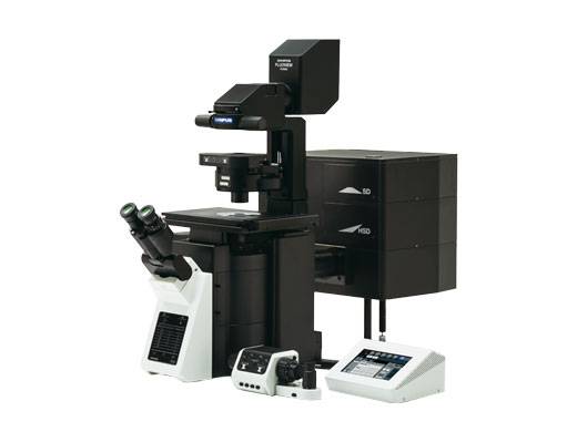
The Olympus FV3000 laser scanning confocal microscope is designed for high-resolution confocal observation of fixed and living cells, point-detection, spectral detection that does variable bandwidth filtering, high efficiency of excitation, 3-D imaging, and time course experiments. This confocal microscope is equipped with 5 diode lasers – 405 nm, 445 nm, 488 nm, 561 nm, and 640 nm. It has high sensitivity and speed capabilities required for live cell and tissue imaging, supports complete workflows from live cell 2D–6D (x,y,λ,z,t,p) imaging through image processing – deconvolution and analysis. It has four spectral confocal detectors with Olympus TruSpectral high-efficiency spectral detection system – 1-100 nm adjustable emission bandwidth with 2 nm spectral resolution, allows users to adjust emission detection for any wavelength between 400 nm and 800 nm. An additional Ir detector has a single red-shifted GaAs PMT with high-sensitivity up to 900 nm, including a 750 nm longpass filter for detection. Sample focusing through oculars via transmitted light or LED epifluorescence. The Olympus IX83 motorized inverted microscope frame has a motorized z motor with 10 nm precision and 3 mm/s speed, motorized revolving objective nosepiece, motorized fluorescence mirror turret, ultrasonic high-speed and high-precision linear-encoded automated stage with software control, and motorized light path selection. Stage sample holders included are the universal slide/petri dish holder, multiwell plate holder, and 4 slide holder. The Bioptechs Delta T5™ heated stage allows for live cell imaging, on an open dish system.
Capabilities
- Objectives:
- Olympus UPLXAPO 4x objective, NA 0.16
- Olympus UPLXAPO 10x objective, NA 0.40
- Olympus UMPlanFL N 10x water objective, NA 0.30
- Olympus UPlanSApo 20x objective, NA 0.75
- Olympus UCPLFLN 20x objective, NA 0.7
- Olympus UPlanSApo 30x silicone immersion objective, NA 1.05
- Olympus UPlanApo 40x oil immersion objective, NA 1.00 Iris
- Olympus UPLXAPO 60x oil immersion Objective, NA 1.42
- Olympus LUMPlanFL N 60x water objective, NA 1.00
- Olympus UPlanApo 60x oil high resolution, NA 1.50
- Advanced techniques:
- Lambda scanning
- Bright Z
- Trufocus/Z-drift compensation system
- Macro to Micro Imaging/Tiling/Mosaic Image Stitching
- Galvanometer and Resonant Scanners (resonance scanner capable of 30 fps@ 512x512 and up to 438 fps @ 512x32 with scanning field number of 18 and continuously adjustable room ratio of 1.0x to 8.0x)
- Advanced software image editing (ex: Cell Sens, deconvolution, cell counts)
Related Equipment
Location: Supple 171
Manager: Dr. Casey Smith, casey.smith@txstate.edu
Model: Olympus FV3000 Laser Scanning Confocal Microscope
Funded by: The Materials Applications Research Center (MARC)